New way of picturing proteins using graphene

Updated: Oct 7, 2018
Until now, the only images of proteins we had were only averaged results of scanning millions of molecules. But in 2015, Jean-Nicolas Longchamp of the University of Zurich, Switzerland and his team came up with a new technique using graphene that gives us a much better idea of protein structure.
Blurry images
There are three main challenges in this technique: how to isolate single proteins, how to keep them fixed in one place long enough to create an image and finally, how not to damage them in the process of scanning. Previous techniques, such as X-ray crystallography or cryo-electron microscopy have failed to meet the third requirement - each attempt at scanning the protein damaged it so much, that it couldn't be scanned again. That is why they took pictures of several molecules and then created an average. The problem with this technique? We get only a very general idea of the protein's shape. (1, 2)
Protein deposition
Unlike in X-ray crystallography, where proteins have to be arranged into crystals before analysing, Longchamp's technique needs a single molecule of an individual protein or a protein complex. This is achieved by an elecrospray, which accelerates the particles and then slows them down before depositing them on a graphene sheet, to ensure a soft landing. (1, 3)

Low-energy electron holography
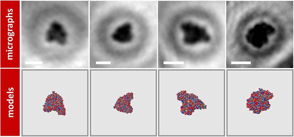
The sheet is then scanned with a low-energy electron holography setup. A coherent beam of electrons is released from an electron point source (EPS). Part of the electron wave is elastically scattered off the object, whereas the unscattered part of the wave represents the reference wave. At a distant detector, the pattern resulting from the interference of these two wave fronts, is recorded, and an image is produced. (3)
The reason for using low-energy electrons are that they don't damage the structure of the protein whilst uncovering fine details of its structure. The short wavelength of the electrons also allows them to pass through the graphene sheet, so that they can be recorded by the detector. (1)

Results

The future of protein imaging
A deeper understanding of protein structure will be useful to many areas of science, particularly structural biology. It could even be the first step to finding cures to diseases that are connected to protein structure, like Alzheimer's, Parkinson's, Creutzfeldt-Jakob disease and Transthyretin amyloidosis. Longchamp's latest research was particularly on imaging amyloid fibrils, that are often the cause to some of these diseases. (2, 4)
As we see, finding new ways of utilising this higher level of protein imaging is certainly exciting for researchers, but it will also most probably give insight into the way many diseases work.
References:
1 Longchamp J. N. (2015). How to image a single protein. Available: https://goo.gl/Vod3fu. Last accessed 7th Oct 2018.
2 Aron, J. (2016). First ever pictures of single proteins thanks to graphene sheet. Available: https://goo.gl/Qjxmrf. Last accessed 7th Oct 2018.
3 Longchamp J. N., Rauschenbach S., Abb S., Escher C., Latychevskaia T., Kern K., Fink H. W. (2017). Imaging proteins at the single-molecule level. Available: https://goo.gl/W12X4V. Last accessed 7th Oct 2018.
4 Coghlan, A. (2015). Universal plaque-busting drug could treat various brain diseases. Available: https://goo.gl/Y6TQsZ. Last accessed 7th Oct 201


Well done Anna. That is an excellent summary and I especially like te diagrams and the way you have given clear references at the end.